Decoding The Eye: A Complete Information To The Anatomical Chart
Decoding the Eye: A Complete Information to the Anatomical Chart
Associated Articles: Decoding the Eye: A Complete Information to the Anatomical Chart
Introduction
With enthusiasm, let’s navigate by the intriguing matter associated to Decoding the Eye: A Complete Information to the Anatomical Chart. Let’s weave fascinating info and provide contemporary views to the readers.
Desk of Content material
Decoding the Eye: A Complete Information to the Anatomical Chart

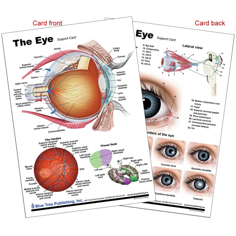
The human eye, a marvel of organic engineering, is chargeable for our sense of sight, permitting us to understand the colourful world round us. Understanding its intricate construction is essential for appreciating its performance and comprehending the assorted circumstances that may have an effect on it. An anatomical chart of the attention supplies a visible roadmap to this advanced organ, highlighting its quite a few elements and their interrelationships. This text delves into the important thing options depicted on a typical eye chart, explaining their roles and significance in sustaining clear and wholesome imaginative and prescient.
I. The Outer Layer: Safety and Refraction
The outermost layer of the attention, chargeable for safety and preliminary mild refraction, includes three foremost buildings:
-
Sclera: This powerful, white, fibrous layer kinds nearly all of the eyeball’s outer floor. Its main perform is to take care of the attention’s form and shield its delicate inside buildings from damage. The sclera’s rigidity is essential for stopping deformation and sustaining the exact curvature crucial for correct picture formation. The seen portion of the sclera is the "white of the attention."
-
Cornea: Located on the entrance of the attention, the cornea is a clear, dome-shaped construction that kinds the anteriormost a part of the attention’s outer layer. In contrast to the sclera, the cornea is avascular (missing blood vessels), relying as a substitute on diffusion from surrounding tissues for oxygen and nutrient provide. This avascularity ensures transparency, essential for permitting mild to go by unimpeded. The cornea performs a major function in refracting (bending) mild rays, contributing considerably to the attention’s focusing energy. Its clean, curved floor is important for correct picture formation on the retina. Corneal irregularities, reminiscent of astigmatism, can considerably impair imaginative and prescient.
-
Conjunctiva: This skinny, clear mucous membrane strains the inside floor of the eyelids and covers the sclera. It acts as a protecting barrier, lubricating the attention floor and offering a protection in opposition to an infection. The conjunctiva’s wealthy blood provide helps to nourish the sclera and aids within the removing of international particles. Irritation of the conjunctiva (conjunctivitis, or "pink eye") is a typical eye ailment.
II. The Center Layer: Vascular Provide and Lodging
The center layer, or uvea, is a vascular layer chargeable for nourishing the attention’s buildings and enjoying an important function in focusing. It consists of three elements:
-
Choroid: This extremely vascular layer lies beneath the sclera, offering oxygen and vitamins to the outer layers of the retina. Its wealthy blood provide is important for supporting the metabolic calls for of the photoreceptor cells. The choroid’s darkish pigmentation helps to soak up stray mild, stopping inside reflections that would blur imaginative and prescient.
-
Ciliary Physique: Situated on the junction of the iris and choroid, the ciliary physique is a ring-shaped construction containing the ciliary muscle and processes. The ciliary muscle controls the form of the lens, permitting the attention to give attention to objects at various distances (lodging). The ciliary processes secrete aqueous humor, the fluid that fills the anterior chamber of the attention.
-
Iris: This coloured, round construction lies behind the cornea and in entrance of the lens. The iris incorporates two units of muscular tissues: the sphincter pupillae, which constricts the pupil, and the dilator pupillae, which dilates it. The pupil, the central opening within the iris, regulates the quantity of sunshine coming into the attention. The dimensions of the pupil adjusts routinely in response to modifications in mild depth, defending the retina from injury by extreme mild and optimizing imaginative and prescient in low-light circumstances.
III. The Interior Layer: Photoreception and Picture Formation
The innermost layer of the attention is the retina, the light-sensitive tissue chargeable for changing mild into electrical alerts which might be transmitted to the mind. The retina’s construction is advanced, containing varied cell varieties:
-
Photoreceptor Cells: These specialised cells are chargeable for detecting mild. There are two foremost varieties: rods, that are delicate to low mild ranges and chargeable for night time imaginative and prescient, and cones, that are chargeable for colour imaginative and prescient and visible acuity in vibrant mild. Cones are concentrated within the macula, a small space close to the middle of the retina.
-
Macula: This specialised area of the retina is chargeable for sharp, central imaginative and prescient. At its middle lies the fovea, a tiny pit containing the best focus of cones, offering the best visible acuity.
-
Optic Disc (Blind Spot): This space of the retina lacks photoreceptor cells, because it’s the place the optic nerve exits the attention. Consequently, it creates a small blind spot in our visible area, which our mind usually compensates for.
-
Optic Nerve: This nerve carries {the electrical} alerts generated by the photoreceptor cells to the mind, the place they’re interpreted as photos.
IV. The Lens: Focusing Gentle onto the Retina
The lens, a clear, biconvex construction situated behind the iris, performs an important function in focusing mild onto the retina. Its flexibility, managed by the ciliary muscle, permits the attention to accommodate for objects at totally different distances. As we age, the lens loses its elasticity, resulting in presbyopia, a situation characterised by problem specializing in close to objects. Cataracts, a clouding of the lens, can even impair imaginative and prescient.
V. The Aqueous and Vitreous Humors: Sustaining Intraocular Stress and Form
The attention incorporates two fluids that preserve its form and intraocular strain:
-
Aqueous Humor: This clear, watery fluid fills the anterior chamber of the attention, the area between the cornea and the lens. It’s repeatedly produced by the ciliary physique and drains by a drainage system. Imbalances within the manufacturing and drainage of aqueous humor can result in glaucoma, a situation characterised by elevated intraocular strain that may injury the optic nerve.
-
Vitreous Humor: This clear, gel-like substance fills the posterior chamber of the attention, the area between the lens and the retina. It helps to take care of the attention’s form and help the retina. With age, the vitreous humor can shrink and detach from the retina, doubtlessly resulting in retinal tears or detachments.
VI. Extraocular Muscle mass and Eyelids:
The attention’s motion is managed by six extraocular muscular tissues, which permit for exact and coordinated eye actions. These muscular tissues are essential for binocular imaginative and prescient (utilizing each eyes collectively to understand depth). The eyelids shield the attention from international our bodies and extreme mild. The lacrimal gland, situated within the higher outer nook of the orbit, produces tears that lubricate and shield the attention floor.
VII. Scientific Significance of Understanding the Anatomical Chart:
A radical understanding of the attention’s anatomy is important for diagnosing and treating a variety of ophthalmic circumstances. The anatomical chart serves as a helpful instrument for:
-
Figuring out the placement of pathology: Figuring out the exact location of a lesion or abnormality throughout the eye is essential for correct analysis and remedy planning.
-
Understanding the pathophysiology of eye illnesses: Many eye illnesses contain particular anatomical buildings. For instance, glaucoma impacts the optic nerve and drainage system, whereas macular degeneration impacts the macula.
-
Guiding surgical procedures: Surgeons use anatomical charts to plan and execute surgical procedures, reminiscent of cataract surgical procedure, retinal detachment restore, and glaucoma surgical procedure.
-
Affected person schooling: Anatomical charts may help sufferers perceive their eye circumstances and remedy choices.
In conclusion, the anatomical chart of the attention supplies a visible illustration of this outstanding organ’s advanced construction and performance. By understanding the person elements and their interrelationships, we will respect the intricate mechanisms that allow us to see the world round us. This data is essential not just for appreciating the great thing about the attention’s design but in addition for diagnosing, treating, and stopping a variety of eye illnesses. The chart serves as a elementary instrument for ophthalmologists, optometrists, researchers, and anybody looking for a deeper understanding of this very important sensory organ.


Closure
Thus, we hope this text has offered helpful insights into Decoding the Eye: A Complete Information to the Anatomical Chart. We respect your consideration to our article. See you in our subsequent article!